TheIncredibleIncel
Veteran
★★★
- Joined
- Jun 17, 2019
- Posts
- 1,216
Source
Prenatal testosterone may have a powerful masculinizing effect on postnatal physical characteristics. However, no study has directly tested this hypothesis. Here, we report a 20-year follow-up study that measured testosterone concentrations from the umbilical cord blood of 97 male and 86 female newborns, and procured three-dimensional facial images on these participants in adulthood (range: 21–24 years). Twenty-three Euclidean and geodesic distances were measured from the facial images and an algorithm identified a set of six distances that most effectively distinguished adult males from females. From these distances, a ‘gender score’ was calculated for each face, indicating the degree of masculinity or femininity. Higher cord testosterone levels were associated with masculinized facial features when males and females were analysed together (n = 183; r = −0.59), as well as when males (n = 86; r = −0.55) and females (n = 97; r = −0.48) were examined separately (p-values < 0.001). The relationships remained significant and substantial after adjusting for potentially confounding variables. Adult circulating testosterone concentrations were available for males but showed no statistically significant relationship with gendered facial morphology (n = 85, r = 0.01, p = 0.93). This study provides the first direct evidence of a link between prenatal testosterone exposure and human facial structure.
Keywords: face shape, testosterone, hormones, prenatal, sexual dimorphism
Go to:
1. Background
The human face is sexually dimorphic, with the average male face differing from the average female face in the size and shape of, and distance between, the jaws, lips, eyes, nose and cheekbones [1,2]. Even within sex, there are considerable variations in these dimensions, leading to individuals appearing more or less feminine or masculine than the prototypical gendered face. While the origin of this variability remains unclear, there has been significant interest in the influence of the most abundant androgen, testosterone, in the development of face structure.
Genetic sex is determined at conception, but gonadal hormones play a vital role in the differentiation of male and female phenotypes throughout human development [3]. To date, studies investigating the influence of testosterone exposure on facial morphology have focused predominantly on hormone exposure during adolescence. Testosterone levels surge during puberty, and concentrations are 20–30-fold higher in males than females [4], which has been postulated to explain the contemporaneous increased sexual dimorphism in facial appearance. For example, among 12–18-year-olds, a positive correlation is present between the concentration of testosterone in saliva and the dimensions of several male-typical facial characteristics, such as a broader forehead, chin, jaw and nose [5]. Furthermore, the administration of testosterone to adolescents with delayed puberty accelerates craniofacial growth in these same features [6]. Experimental studies of adults have also revealed a link between contemporaneous testosterone levels and subjective ratings of facial masculinity [7], as well as more objective measurements, such as increased facial width-to-height ratio (fWHR) [8].
However, there is accumulating evidence that the influence of androgens on the development of secondary sex characteristics begins much earlier in life. Biosynthesis of testosterone commences at approximately nine weeks gestation. While testosterone is made in the adrenal cortex and ovary of females, it is produced in far greater amounts by the Leydig cells of the testes in males, and a sex difference in prenatal testosterone level exposure emerges during the second trimester [9] and persists until birth [10]. Studies of non-human mammals have found that exposure to supraphysiological levels of testosterone has a masculinizing effect on physical development, in the form of increased anogenital distance, reduced nipple number, a smaller vaginal opening and increased body growth [11–14]. Sex differences in facial morphology are apparent in six-month-old infants [15], and increase steadily across childhood [16]. While testosterone surges and associated growth spurts during puberty exaggerate existing sex differences in face structure, it has been postulated that the pre-existing sexual dimorphism in face structure is related to differences in prenatal testosterone exposure [17].
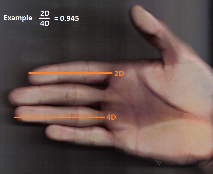
Investigating the proposed link between testosterone and face development in humans has been hampered by two methodological challenges. The first has been the valid and reliable measurement of prenatal testosterone exposure. As it is unethical to manipulate hormone concentrations in the human fetus, the majority of clinical research has used surrogate measures of the prenatal hormone environment, such as the ratio of the second digit to the fourth digit (2D : 4D) [18] and the observation of fetuses exposed to aberrant hormone environments in utero (e.g. females with congenital adrenal hyperplasia) [19,20]. However, limitations of these approaches have been widely documented, with reports of poor correlations between 2D : 4D and more direct measures of the prenatal hormone environment [21,22], as well as concerns about the extent to which data from clinical populations can be extrapolated to the general population.
An alternative approach for sampling the prenatal androgen environment is investigating the hormonal milieu of umbilical cord blood. Cord blood can be obtained at delivery in normal pregnancy, and therefore randomly selected participant samples are more likely to be representative of the general population. Several studies have reported higher testosterone levels in cord blood samples from male versus female fetuses [10,23], and thus these samples are thought to reflect fetal circulation during late gestation. A limitation of this approach is that testosterone levels in cord blood may not reflect concentrations during the first and second trimester, in particular gestational weeks 8–24, which has traditionally been regarded as a ‘sensitive period’ for the maximal effects of sex steroids on human development [24]. However, there is increasing recognition that there may be multiple sensitive periods, and animal studies have found that postnatal development may be affected by hormones at different times throughout prenatal development [25]. Human studies have also started to link sex steroids from umbilical cord blood to a range of childhood behaviours, including language development [26,27], internalizing and externalizing behaviours [28], and spatial abilities [29]. To date, no studies have examined the relationship between cord blood testosterone and human physical development.
The second methodological challenge has been the accurate measurement of facial shape. Previous studies have focused on either subjective masculinity ratings of two-dimensional (2D) facial stimuli [30], or measurements between standardized landmarks on 2D photographs [17,31–34]. Distances based on 2D photographs are unreliable as they change with the camera distance, angle and optics. Some researchers have used three-dimensional (3D) models to calculate Euclidean (linear) distances between anatomical landmarks. However, the use of these measurement techniques may miss important information since Euclidean distances ‘cut through’ the facial features and thus do not take the underlying morphology into account. For example, calculation of some characteristics of facial dimorphism using these distances does not correlate well with human perceptual measurements [35]. In contrast, geodesic (3D surface) distances are known to model the 3D facial structure in a better way as compared with the linear distances. For example, one study has reported that objective scores for masculinity calculated from 3D facial images correlate more highly with subjective (perceptual) masculinity scores if the objective scores are based on Euclidean and geodesic distances, rather than on Euclidean distances alone [35].
The current study represents the first to investigate the potential link between prenatal testosterone exposure and objective measurements of 3D facial masculinity in adults. The current study spanned a 25-year period, in which umbilical cord blood was collected at birth (1989–1991) and facial morphology was examined on the same participants in early adulthood (2012–2014). A total of 97 males and 86 females (all Caucasian) had both cord blood and adult facial morphology data available, and thus provided the data for the current study. Blood collected in early adulthood was available for the male participants only, and testosterone concentrations were measured in these samples. The 2D : 4D measurements from both hands of the adult participants were also procured and included in the analyses.


